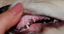
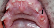
 Dr. Leon has been a Veterinary Specialist for 10 years and is the co-owner of VetDent the first network of Latin American Veterinary Dentists. He has participated in the research group Prof. Gioso (USP) in the laboratory of Comparative Ondontology (LOC) since it’s creation. In 2004, together with Dr. Alexandre Wenceslas, he created the first mobile service for Veterinary Dental services of Brazil. He is also the coordinator of the Center for Training and Education in Dentistry Veterinary CTEOV – participating in the education of many fellow vets who will offer the same quality service in the future. Dr. Leon's website: Dentista Vet. Borzoi International thanks Dr. Leon for contributing this important article to our site and taking exceptional care of Solange Mikhail's Borzoi Yuri as well! My Puppy Broke His Tooth! Cause: Puppies like chewing and Borzoi puppies are no exception. During this phase your puppy can break the deciduous (primary) tooth. A fracture can occur from chewing bones, rocks, metal, toys, any other hard material. Why the broken tooth has to be removed? You may think that because the tooth is "just" a puppy tooth you can wait until if falls out, but that can be a costly and unwise decision. A fractured deciduous tooth is painful and quickly becomes infected. This infection can cause a draining tract, osteomyelitis, or damage to the permanent tooth. Therefore, a "wait and see" choice is not an option. The pulp exposed contains nerve endings and is very painful for the puppy, which can cause loss of appetitite and be harmful to their proper development during a borzoi's fast-growing puppy stage. Therefore, a fractured deciduous tooth with pulp exposure requires extraction therapy as soon as possible Who can treat it? If your puppy breaks a baby tooth it should be carefully evaluated by a veterinarian specializing in canine dentistry. This professional will have appropriate instruments and knowledge to determine the best treatment for the fractured tooth through physical and radiographic evaluations. The tooth layer that is compromised and the age of the animal should be considered. The tooth: The tooth has three layers. The outside layer is a thin layer called the enamel. The second layer consists of a hard substance called dentin. The inside of the tooth is called the dental pulp, which is made up of arteries, veins, nerves and connective tissue. Once the pulp is exposed, it is very painful for the dog and oral bacteria can infect and kill the pulp as well as damage the surrounding tissues. Usually, for a fracture that involves pulp exposure in puppies, the treatment of choice is extraction. In adult dogs the treatment is different: everything is made to preserve the tooth (pulpectomy, root canal therapy, restorative treatment with metal crowns) – as they are the permanent teeth. 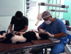 Evaluation: During the evaluation it is necessary that the patient is under general anesthesia or sedated to avoid movement. It is usually a quick procedure, but because Borzoi are very sensitive to anesthesia, this topic should be carefully discussed with your veterinarian. The intra-oral radiographs provide superior quality for examination of the fractured tooth with less radiation risk than standard-sized veterinary radiographs. Pre-extraction x-rays are essential to identify the location of the permanent tooth. A dental radiograph is helpful to determine the position of the permanent tooth as well as the extension of the fracture. The physical evaluation can be performed on the fractured tooth using a dental explorer and a periodontal probe. It can detect loose fragments, cracks, multiple fracture planes, separation between dentin and enamel and exposed pulp canals. The area below the gumline can also be checked. The extraction: A fractured deciduous tooth should be carefully extracted with proper instruments. This requires skill and knowledge of dental anatomy. The procedure is delicate, since the permanent teeth (in the process of forming) are really close and can be damaged with any extra pressure or trauma in the region. The goal is to extract the tooth and its entire root without causing damage to the developing permanent tooth. A general anesthesia is crucial for a successful procedure. Any small twist can break the deciduous tooth at the moment of the extraction, as it is a very thin and fragile structure. If that happens, it will involve more work and time to remove it and will need a larger incision. 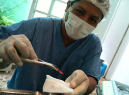 Fig. 4 Fig. 4 Fig. 4: Successful extraction of the deciduous fractured tooth 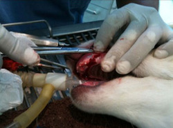 Fig. 5 Fig. 5 Extraction will result in a large hole where the tooth used to be, and necessitates that the tissues be sutured without tension (with absorbable stitches) to prevent food and debris from getting trapped in the wound. To the left in Fig. 5 the suture is being performed. Preventive care: While it is hard to change the behavior of puppies, try to avoid their chewing hard things like rocks, gates or any metal or iron housing. Do not buy hard bones as a treats. Rawhide or rubber bones are preferable. A collection of soft toys can keep your Borzoi busy enough not to pay attention to the forbidden things. 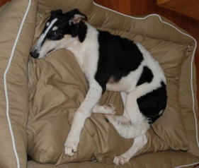 Fig. 6: Yuri at home recovering in his comfy bed! We thank Dr. Leon for taking such good care of Yuri and allowing Solange to be in the operating room and take pictures of Yuri’s procedure. Thanks go to Solange Mikhail for her help in translating the article for the Borzoi International Blog.
5 Comments
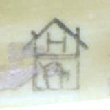
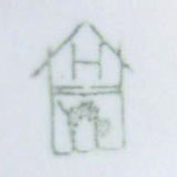
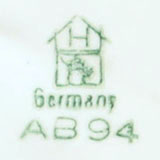
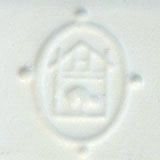
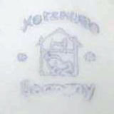
 Many Borzoi enthusiasts and collectors are familiar with the fine Art Deco era porcelains produced by Hertwig and Company of Germany. Katzhutte, once referred to as “Poor Man’s Goldscheider” has now become a sought after class of art with rare pieces becoming quite valuable. Katzhutte means cat house in German and was started back in the 1800’s in Thuringia, Germany. Pieces produced at this factory carry the green or blue cat in house marking. The factory was started in 1864 when Nikolas Beyerman and Ernst Fredrich Hertwig acquired the “UntereEisenhammer” a rather old and run down iron works on the bank of the Schwarzariver in Germany. There was good clay there and an abundance of cheap skilled labor. By 1890 there were 900 people employed by Hertwig, 300 in the factory on production and 600 home workers who made the clothes and bodies of the dolls. Puppets and Dolls were what Hertwig was most famous for. They also produced animals, birds and utilitarian kitchenware. 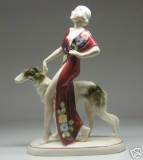 In 1958 the East German State took the factory over and the company was very unprofitable. In 1983 the state needed hard currency, so they raided the factory, museum and showrooms where copies of each piece produced by the Hertwig factory since 1864 were held, and they expropriated them all. It is estimated that some 15,000 items disappeared and were converted into hard currency in the West. So the records were lost. The factory continued to decline and in 1995, it finally closed. During their peak, the factory produced many lovely Borzoi pieces among their collections.The works during this time would often include lovely ladies with Borzoi in an Art Deco style. There were also many lovely Borzoi pieces created of Borzoi on their own in running, standing and sitting positions.
What is DM?
Degenerative myelopathy is a progressive disease of the spinal cord in older dogs. The disease has an insidious onset typically between 8 and 14 years of age. It begins with a loss of coordination (ataxia) in the hind limbs. The affected dog will wobble when walking, knuckle over or drag the feet. This can first occur in one hind limb and then affect the other. As the disease progresses, the limbs become weak and the dog begins to buckle and has difficulty standing. The weakness gets progressively worse until the dog is unable to walk. The clinical course can range from 6 months to 1 year before dogs become paraplegic. If signs progress for a longer period of time, loss of urinary and fecal continence may occur and eventually weakness will develop in the front limbs. Another key feature of DM is that it is not a painful disease. What causes degenerative myelopathy? Degenerative myelopathy begins with the spinal cord in the thoracic (chest) region. If we look under the microscope at that area of the cord from a dog that has died from DM, we see degeneration of the white matter of the spinal cord. The white matter contains fibers that transmit movement commands from the brain to the limbs and sensory information from the limbs to the brain. This degeneration consists of both demyelination (stripping away the insulation of these fibers) and axonal loss (loss of the actual fibers), and interferes with the communication between the brain and limbs. Recent research has identified a mutation in a gene that confers a greatly increased risk of developing the disease. How is DM clinically diagnosed? Degenerative myelopathy is a diagnosis of elimination. We look for other causes of the weakness using diagnostic tests like myelography and MRI. When we have ruled them out, we end up with a presumptive diagnosis of DM. The only way to confirm the diagnosis is to examine the spinal cord under the microscope when a necropsy (autopsy) is performed. There are degenerative changes in the spinal cord characteristic for DM and not typical for some other spinal cord disease. How do we treat degenerative myelopathy? There are no treatments that have been clearly shown to stop or slow progression of DM. Although there are a number of approaches that have been tried or recommended on the internet, no scientific evidence exists that they work. The outlook for a dog with DM is still grave. The discovery of a gene that identifies dogs at risk for developing degenerative myelopathy could pave the way for therapeutic trials to prevent the disease from developing. Meanwhile, the quality of life of an affected dog can be improved by measures such as good nursing care, physical rehabilitation, pressure sore prevention, monitoring for urinary infections, and ways to increase mobility through use of harnesses and carts. Testing for degenerative myelopathy: The collaborative efforts of Dr Joan Coates and Dr Gary Johnson and associates at the University of Missouri and Dr Kirsten Lindblad-Toh and Dr Claire Wade and associates at the Broad Institute at MIT/Harvard have resulted in identification of a mutation that is a major risk factor for the development of Degenerative Myelopathy in many breeds of dogs. The DNA test for DM is now available through the Orthopedic Foundation for Animals (OFA). This test clearly identifies dogs that are clear (have 2 normal copies of the gene), those who are carriers (have one normal copy of the gene and one mutated copy of the gene), and those who are at much higher risk for developing DM (have 2 mutated copies of the gene). However, having two mutated copies of the gene does not necessarily result in disease. Dogs that have clinical signs or a presumptive diagnosis of DM have tested as genetically affected. A relatively high percentage of dogs in several breeds (including Boxers, Chesapeake Bay Retrievers. Pembroke Welsh Corgis, and Rhodesian Ridgebacks) have the predisposing mutation. It is important to note that there are a large number of dogs that have tested as genetically affected, but are reported as clinically normal by their owners. Information in the RESEARCH section of this website outlines continued and ongoing research that seeks to understand what triggers development of clinical symptoms in some, but not all dogs at risk. Understanding the DNA test for degenerative myelopathy: We have discovered a gene which is a major risk factor for degenerative myelopathy (DM). In that gene, the DNA occurs in two possible forms (or alleles). The “G” allele is the predominant form in dog breeds in which DM seldom or never occurs; you can think of it as the “Good” allele. The “A” allele is more frequent in dog breeds for which DM is a common problem; you can think of it as the “Affected” allele. Summary: “A” allele is associated with DM; “G” allele is not associated with DM. Since an individual dog inherits two alleles (one from the sire and one from the dam) there are three possible test results: two “A” alleles; one “A” and one “G” allele; and, two “G” alleles. Summary: Test results can be A/A, A/G, or G/G. In the seven breeds we studied so far (Boxer, Chesapeake Bay Retriever, German Shepherd Dog, Pembroke Welsh Corgi, Cardigan Welsh Corgi, Rhodesian Ridgeback, and Standard Poodle), dogs with test results of A/G and G/G have never been confirmed to have DM. Essentially all dogs with DM have the A/A test result. Nonetheless, many of the dogs with an A/A test result have not shown clinical symptoms of DM. Dogs with DM can begin showing signs of disease at 8 years of age, but some do not show symptoms until they are as old as 15 years of age. Thus, some of the dogs who have tested A/A and are now normal may still develop signs of DM as they age. We have, however, found a few 15-year-old dogs that tested A/A and are not showing the clinical symptoms of DM. Unfortunately, at this point we do not have a good estimate of what percent of the dogs with the A/A test result will develop DM within their lifespan. Summary: Dogs that test A/G or G/G are very unlikely to develop DM. Dogs that test A/A are much more likely to develop DM. Our research will now focus on how many A/A dogs can survive to old age without developing DM and why. The “A” allele is very common in some breeds. In these breeds, an overly aggressive breeding program to eliminate the dogs testing A/A or A/G might be devastating to the breed as a whole because it would eliminate a large fraction of the high quality dogs that would otherwise contribute desirable qualities to the breed. Nonetheless, DM should be taken seriously. It is a fatal disease with devastating consequences for the dogs and a very unpleasant experience for the owners who care for them. Thus, a realistic approach when considering which dogs to select for breeding would be to consider dogs with the A/A or A/G test result to have a fault, just as a poor top-line or imperfect gait would be considered faults. Dogs that test A/A should be considered to have a worse fault than those that test A/G. Dog breeders could then continue to do what conscientious breeders have always done: make their selections for breeding stock in light of all of the dogs’ good points and all of the dogs’ faults. Using this approach over many generations should substantially reduce the prevalence of DM while continuing to maintain or improve those qualities that have contributed to the various dog breeds. Summary: We recommend that dog breeders take into consideration the DM test results as they plan their breeding programs; however, they should not over-emphasize this test result. Instead, the test result is one factor among many in a balanced breeding program. This article has been approved to publish by the University of Missouri staff. For additional info please go to the following websites: www.CanineGeneticDiseases.net www.OFFA.org |
A New Article Each Quarter!
Archives
July 2014
Categories
All
|

 RSS Feed
